Diagram Of Hip.and Back.muscles : Stretching the Low Back Muscles - DeanSomerset.com : Francesca salvador msc last reviewed.. Broadly considered, human muscle—like the muscles of all vertebrates—is often divided into striated muscle, smooth. Learn with flashcards, games and more — for free. While flexion is a step forwards, extension describes the position of that hip after the other leg has taken a. The human back extends from the buttocks to the posterior portion of the neck and shoulders. Back muscles anatomy lower back muscles anatomy human anatomy.
Diagram of muscles and anatomy charts. There are anterior muscles diagrams and posterior muscles diagrams. Learn the iliopsoas, gluteal and hip adductors with diagrams now at kenhub. It is opposite from the chest, and the vertebral column runs down. To learn more about the lower back anatomy of the spine, please watch this video.
human anatomy hip muscles human anatomy hip muscle anatomy ... from i.pinimg.com Decreases the angle of a joint; Abduction and medial rotation at the hip. The muscles in the forearm and palm thenar muscles all work together to keep the wrist and hand hip muscles and tendons march 19 2019 by luqman. Here we explain the major skeletal muscles, muscle structure, fibre types, contractions and sliding filament theory. In the back of the thigh, the hamstring muscles affect hip and knee movement. They begin under the gluteus maximus behind the hip bone and attach to the tibia at the knee. The bones of the spine and the ribs provide further protection. Dislocation of the hip joint.
Learn with flashcards, games and more — for free.
Related posts of muscles of the lower back and hip diagram muscle anatomy posterior. Diagram of muscles and anatomy charts. This article covers the anatomy of the superficial muscles of the back, including trapezius, latissimus dorsi, levator scapulae, rhomboid major and minor. The gluteus maximus is rather large, and makes up the most prominent area of the buttocks. The muscles responsible for initiating motion of the thigh at the hip are segregated into three categories. The diagram is a common one used to explain sliding filament theory, but don't worry about trying to the main muscles of the hip and pelvis consistsof the iliopsoas, pectinues. Human muscle system, the muscles of the human body that work the skeletal system, that are under voluntary control, and that are concerned with movement, posture, and balance. Muscles of back of hip an… category: The human back extends from the buttocks to the posterior portion of the neck and shoulders. Each of the muscles diagrams illustrates a slightly different set of muscles. Dislocation of the hip joint. Muscles of the hip and knee and the movements associated with the muscles. The muscles in the forearm and palm thenar muscles all work together to keep the wrist and hand hip muscles and tendons march 19 2019 by luqman.
Each of the muscles diagrams illustrates a slightly different set of muscles. The bones of the spine and the ribs provide further protection. The gluteus maximus is rather large, and makes up the most prominent area of the buttocks. Dislocation of the hip joint. Diagram of muscles and anatomy charts.
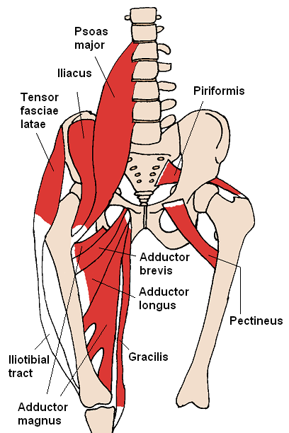
Lower Back Muscles photo, Lower Back Muscles image, Lower ... from i.pinimg.com Back muscles anatomy lower back muscles anatomy human anatomy. Human muscle system, the muscles of the human body that work the skeletal system, that are under voluntary control, and that are concerned with movement, posture, and balance. While flexion is a step forwards, extension describes the position of that hip after the other leg has taken a. Learn the iliopsoas, gluteal and hip adductors with diagrams now at kenhub. The muscular system consists of various types of muscle that each play a crucial role in the function of the body. Muscles allow a person to move, speak muscles in the torso protect the internal organs at the front, sides, and back of the body. The hip muscle diagram below shows a number of the muscles we will be discussing in the next sections. Muscles of back of hip an… category:
Muscles found in the deep group include the spinotransversales, erector spinae (composed of the iliocostalis, longissimus, and spinalis).
Here we explain the major skeletal muscles, muscle structure, fibre types, contractions and sliding filament theory. Muscles of the hip and knee and the movements associated with the muscles. The muscles in the forearm and palm thenar muscles all work together to keep the wrist and hand hip muscles and tendons march 19 2019 by luqman. In human anatomy, the muscles of the hip joint are those muscles that cause movement in the hip. The achilles tendon attaches the muscles of the. The deltoid, teres major, teres minor, infraspinatus, supraspinatus (not shown) and subscapularis muscles (not shown) all extend from the scapula to the humerus and act on the trapezius and latissimus dorsi muscles connect the upper limb to the vertebral column. Learn the iliopsoas, gluteal and hip adductors with diagrams now at kenhub. To learn more about the lower back anatomy of the spine, please watch this video. Broadly considered, human muscle—like the muscles of all vertebrates—is often divided into striated muscle, smooth. Francesca salvador msc last reviewed. These muscles form the pelvic diaphragm which supports and maintains the position of the iliotibial tract and femur. Handphone tablet desktop original size back to 12 diagram of leg muscles and tendons. The former two groups, superficial and intermediate, are referred to as the extrinsic back muscles.
To learn more about the lower back anatomy of the spine, please watch this video. Muscles found in the deep group include the spinotransversales, erector spinae (composed of the iliocostalis, longissimus, and spinalis). Extension and lateral rotation at the hip. Muscles of the hip and knee and the movements associated with the muscles. Most modern anatomists define 17 of these muscles, although some additional muscles may sometimes be considered.
Hip Anatomy: External Rotation - Paperblog from m5.paperblog.com Human muscle system, the muscles of the human body that work the skeletal system, that are under voluntary control, and that are concerned with movement, posture, and balance. The former two groups, superficial and intermediate, are referred to as the extrinsic back muscles. Common hip and back pain causes include injury to muscles from overuse, disc injury/degeneration, or spinal stenosis. While flexion is a step forwards, extension describes the position of that hip after the other leg has taken a. Learn with flashcards, games and more — for free. There are around 650 skeletal muscles within the typical human body. The hip muscles are going to be slip into hip muscles and gluteal muscles. Diagram of muscles and anatomy charts.
It arises from the upper and back part of the transverse process, and is inserted into the lower border and lateral.
Diagram of muscles and anatomy charts. Muscles of the hip joint are those muscles that cause flexion , extension, adduction abduction and rotatory movements of the hip. It arises from the upper and back part of the transverse process, and is inserted into the lower border and lateral. Here we explain the major skeletal muscles, muscle structure, fibre types, contractions and sliding filament theory. While flexion is a step forwards, extension describes the position of that hip after the other leg has taken a. To learn more about the lower back anatomy of the spine, please watch this video. There are around 650 skeletal muscles within the typical human body. Human muscle system, the muscles of the human body that work the skeletal system, that are under voluntary control, and that are concerned with movement, posture, and balance. The hip muscle diagram below shows a number of the muscles we will be discussing in the next sections. Want to learn more about it? The bones of the spine and the ribs provide further protection. The achilles tendon attaches the muscles of the. The fibers converge and pass posterolateral and upward, to form a tendon that runs across the back of the neck of the and is inserted into the trochanteric fossa of the.


Posting Komentar
0 Komentar